Irina Larina, Baylor College of Medicine, can pinpoint the moment she realized it might just be possible to truly understand the mysterious and largely overlooked world inside women’s fallopian tubes—and their impact on fertility. It was April 2022, and she was watching University of Arizona optical scientist Jennifer Barton explain to a crammed auditorium how her mini endoscopes could detect very early ovarian cancer. Barton had spent years developing submillimeter-diameter falloposcopes tiny enough to peer inside the fallopian tubes, and now her instruments had reached human trials, being guided by surgeons through the uteruses and into the narrow tubes of female volunteers, to collect live data.
Larina herself had been developing 3D optical coherence tomography (OCT) methods with novel algorithms to study reproduction in vivo—but in mice. With her team, she had already tracked embryos moving along mouse fallopian tubes and imaged cilia, hairlike tendrils, on cells lining the tubes, linking her observations to fertility. But sitting in the packed auditorium at a biophotonics congress in Florida, Larina was astonished to learn that falloposcopes were probing similar terrain in women—females whose reproductive tracts are more challenging to reach than in mice.
“Jennifer was capturing these images of cancer cells and tissue in women’s fallopian tubes, and I remember thinking, ‘wow—if only we could do some of our reproduction analysis in females [as well as mice]’,” she says. “It seemed almost impossible at the time, but it got me thinking—and then a few months later Jennifer came to Houston to give a seminar on her work, so we started talking, then brainstorming, and here we are now.”
In summer 2025, Larina and Barton won a five-year, multimillion-dollar National Institutes of Health (NIH) grant to combine falloposcopy and OCT and develop new algorithms, explore the dynamics of cilia in females, and relate their findings to endometriosis. This leading cause of infertility—a condition where cells similar to those in the lining of the womb (uterus) grow in other parts of the body, including the ovaries and fallopian tubes—can damage fallopian tubes and affects more than 10% of women aged 15 to 44. It can remain undetected for years. As Larina puts it: “This subject is under-investigated, under-diagnosed, and tools are rudimental.”

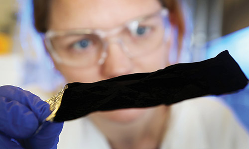
The falloposcope was developed by Jennifer Barton and colleagues at the University of Arizona (UA). Photo credit: Jennifer Barton, University of Arizona
Past studies have revealed that the diminutive fallopian-tube cilia, only 5 to 10 μm in length and some 300 nm in diameter, bend and sway in coordinated, wavelike patterns. However, a few pilot studies have also linked endometriosis to erratic cilia motion, although which is cause and which is effect remains unclear.
So, over the next few years, Barton, Larina, and their research teams will develop and integrate the falloposcope, OCT, and algorithms to identify differences in cilia motion as accurately as possible in humans. They can’t wait for the results. “You know, nobody totally understands how [fallopian tube] cilia work, not even throughout the menstrual cycle of healthy women—so to us, this is a wonderful NIH grant,” says Barton. Still, the anticipated discoveries could never happen without the ingenious developments from each researcher, and the remarkable insights into the complex hidden workings of the fallopian tubes that followed.

Dilara Long, an MD/PhD candidate at UA, assists with the falloposcope. Photo credit: Larina/Barton.
Barton, who was 2024 SPIE President, describes herself as an “engineer and tool builder.” As a biomedical engineering grad student at the University of Texas, Austin, in the 1990s, she was developing high-resolution OCT to image blood vessels live, in the skin, to better diagnose cardiovascular disease. In the early 2000s, a gynecological surgeon suggested Barton explore ovarian cancer. At the time, patients would visit a doctor with minimal symptoms only to find out they had advanced ovarian cancer. The lack of reliable screening methods back then meant women were dying. The five-year survival rate for ovarian cancer in the US was 44%.
“My colleague told me [that] these patients would receive surgery and chemotherapy, be fine for a couple of years, but then the cancer would recur and was almost always deadly,” recalls Barton. “This was so difficult—those couple of years were long enough for [Barton’s colleague] to get to know her patients really well, and then they’d pass with the disease.”
Barton swiftly started to adapt OCT imaging to identify abnormal tissue growth on the ovaries but then spent years trying to figure out why these and other telltale signs of ovarian cancer couldn’t be located on the organs. That is, until 12 years ago, when geneticists discovered that ovarian cancer doesn’t originate in the ovaries; rather, it begins in the fallopian tubes as serous tubal intra-epithelial carcinoma lesions.
For Barton, this fundamental science discovery changed everything. “Cells from these precursor lesions were breaking off the fallopian tubes and floating down to the ovaries or elsewhere in the abdominal cavity, and then growing very quickly,” she says. It changed her team’s engineering approach. “We needed to design and develop miniature endoscopes that could go up though the vagina and uterus, and then through the 1 mm-diameter opening of the fallopian tubes to look at the lesions with high resolution.” Today, Barton’s patented falloposcopes are, quite simply, endoscopes re-imagined. Your everyday endoscope used for, say, a colonoscopy, is a long, thin tube with a small camera and light on the end for seeing inside the body. However, Barton’s miniature versions have swapped the straightforward camera and light for a fiber-bundle comprising thousands of fiber strands, a tapered illumination fiber, and several lenses—all packed into an 800 µm diameter plastic sheath that can be directed into the fallopian tubes via a guide wire.
Upon entering the fallopian tubes, the illumination fiber shines light from the falloposcope tip onto tissue, which reflects and is collected by a small lens at the tip of the fiber bundle. By imaging the other end of the fiber bundle, real-time images of tissue can be seen on a computer screen as the falloposcope travels through the tubes.
But there’s more. Barton’s falloposcopes also contain a single-fiber OCT probe. OCT is based on the analysis of interferometry between light backscattered from a sample and a reference signal. In the falloposcope, the OCT probe emits light from the falloposcope tip that is then focused onto the tissue via lenses. The backscattered light is captured by the probe and the resulting interference patterns are reconstructed to create high resolution cross-sectional images of the fallopian tube wall.
An ingenious part of the entire falloposcopy setup lies in the innovative integration of the different imaging systems from Barton and her team. “When we design our mini endoscopes, we source the parts we need, bring them back to the lab, and do the assembly,” says Barton. “But a lot of our work benefits from technology developments from the companies we work with.” Barton points out that, while the first falloposcopes included a rather rigid 3,000-element fiber bundle, the latest versions use recently developed flexible 9,000-element fiber bundles that can more easily thread their way around the bends of a fallopian tube. “Our first sheaths [surrounding the fiber bundles] were too stiff [to bend around the fallopian tube], but plastic extrusion technology has since gotten better [more flexible],” she says.
Critically, Barton and her team believe their integration of fiber-bundle fluorescence imaging and OCT has, so far, delivered the best-possible view of the fallopian tubes and ovarian cancer’s precursor lesions. Their latest human trials have captured high resolution images of tissue structures in the fallopian tubes and helped them to establish what normal tube tissue should look like.
“We’ve been working on this for so long because we’ve had to wait for many developing technologies, including lasers, fibers, and materials,” she adds. “But things that weren’t possible when I first looked into this really are possible today.”
Just as Barton broke new ground on the once-neglected topic of ovarian cancer, Larina has forged ahead in the almost-forgotten field of female fertility. As she puts it: “With mice, we’ve explored fundamental questions described as fact in books—but when we actually look at the processes, we make discovery after discovery. Why? It’s because the reproduction system is so unexplored.”
Like Barton, Larina knew she wanted to work with OCT, thanks to its ability to resolve tissue structures, such as cilia, at up to 3 mm depths. But as well as imaging tissue detail at these depths, she wanted to study cilia dynamics, and how embryos move along the fallopian tubes, to really get to grips with the biology, so algorithm development began apace.

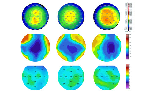
Cilia in human fallopian tubes. Top panel: OCT image of the surface of the fallopian tube, with color-coded mapping of the locations of cilia. Bottom panel: Colored stripes correspond to the phase of beating cilia, showing how their dynamics change over time. Photo credit: Irina Larina.
As a postdoctoral researcher at Baylor College of Medicine in the late 2000s, Larina hadn’t yet started any fertility studies, but instead was looking at embryonic development in mice, and devising algorithms to explore how the heart and vasculature develop. “The heart beats periodically, so we developed many algorithms to help image this periodicity—and these were surprisingly relevant to our [later] studies on cilia dynamics,” she says.
Larina switched to studying the fallopian tubes of mice to better understand fertility about a decade ago. “If something doesn’t work, people tend not to talk about it, and reproductive system disorders are certainly not something women advertise,” she says. “I thought—and still think—that this subject has been avoided by [biomedical] engineers for a long time, but it’s so important as it relates to half the population.”
Larina and colleagues initially developed a high-speed OCT platform for in vivo imaging of fallopian tubes in anaesthetized mice. In their set-up, a laser was directed onto fallopian tube tissue that had been gently pulled through a small slit in the mouse’s back. The backscattered light was captured by a high-speed camera and analyzed to create high resolution depth-resolved 3D images of developing features in the ovary, oocytes, and fallopian tube opening.
More recently, Larina and colleagues have developed OCT systems that rely on small windows 3D printed in their lab and then surgically implanted into the backs of mice. By positioning an OCT imaging probe against the window, they directly view processes within the fallopian tubes, such as cilia motion and embryo transfer from the ovaries to the womb. Critically, they also analyze observations with custom algorithms, including innovative fast Fourier transform-based methods that correlate fluctuations in the pixel intensity of OCT images to the movement of cilia. Their studies have already delivered a few surprises.
During ovulation, for example, fimbriae, fingerlike projections at the opening of the fallopian tubes, were always thought to pick up an ovulated egg and sweep it into the fallopian tube, ready for transfer to the uterus. However, Larina and colleagues noticed that smooth muscles along the fallopian tubes of their mice contract and pump secretions into the tubes, flushing the egg through the passage.
The researchers also reckon these smooth muscles could be responsible for the unexpected commotion they have discovered taking place in mice fallopian tubes. Embryos were always thought to move slowly but surely along the fallopian tubes, gently swept along by the swaying cilia—disrupting this process was also considered to impair fertility. However, the latest observations reveal the fertilized eggs are spinning and careering along these tracts, stopping and restarting probably as the smooth muscles contract and relax. “We’re seeing so much mechanical stimulation—there’s this complexity no one expected,” says Larina.
“We now realize that cilia in fallopian tubes of mice are doing things very differently from what clinicians believed for many, many decades since their discovery,” she adds. “This leaves us questioning the role of these structures in the fallopian tubes of women.”
Clearly, the discoveries in mice could have profound implications for reproductive mechanisms and infertility in women, but can the observations be easily translated to humans? Given Larina and Barton’s latest NIH project to combine their imaging innovations to explore the dynamics of cilia in females, and to relate their findings to endometriosis, we could soon find out.

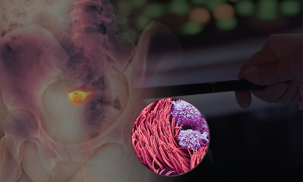
Intravital OCT reveals an embryo—before implantation—travelling through a fallopian tube of a mouse. Photo credit: Irina Larina.
“No one has really looked at human fallopian tubes like this before, so we don’t know exactly what we’re going to find,” says Larina. “But we have already been imaging human fallopian tube samples, and it’s been surprising to what degree these [images] and datasets resemble what we’ve seen in mice.”
As the development and integration of imaging tools begin again to unravel more of the mysteries of the now, not-quite-so-overlooked field of female reproductive health, both Barton and Larina are under no illusion that the next five years of the NIH project will be straightforward. For starters, the falloposcope design needs an overhaul, as the instrument will be imaging groups of swaying cilia and not ovarian cancer lesions. And on top of this, imaging in vivo in humans isn’t easy, a fact Barton knows only too well.
“When you’re in vivo, all of a sudden everything’s moving,” she says. “The living tissue is alive and moving, you’ve got an endoscope that’s moving, you’ve got the physician holding the endoscope that’s moving, and you’ve got the patient that’s moving.”
Given this, the new falloposcope will need to be supremely stable to minimize imaging artefacts that arise as it moves through the turbulent environment within the fallopian tubes. Also, yet-to-be-developed algorithms will have to differentiate between cilia motion and other sources of movement. And that is just the beginning.
“We’ve got to make sure we’re bringing in the right technologies together,” says Larina. “So, we’re looking at lenses, fiber-bundle-density, cameras, and the ways that scans can be performed—all of which depend on what parameters we need to capture, which is something we don’t completely understand yet.”
Still, innovation is underway. For the new falloposcope, Barton and her team are developing proximal helical scanning, which involves attaching a small motor to the end of the OCT fiber that remains outside the human body whilst imaging takes place within the fallopian tubes. As Barton highlights, the motor spins the fiber so surgeons will be able to scan all around the inside of the fallopian tubes in their future searches for endometriosis markers.
“As well as these radial scans, the surgeon can also pull the OCT fiber to take helical scans as it passes through the fallopian tubes,” she says. “This is just really nice.”
Barton’s earlier falloposcope technology is licensed, and come the end of the project, she is hopeful that her technologies will be commercialized and will reach more female patients. “I think there’s finally enough of a critical mass of women in biomedical optics for us to say, ‘we have these tools and we should be applying them to these conditions’—whether that’s normal conditions such as the menopausal transition, or disorders such as endometriosis and infertility,” she says.
Larina concurs and believes it’s high time the world knew more about fallopian tubes and fertility. “Even in reproductive biology, fallopian tubes have been ignored,” she says. “But the fact we have the funds for our research in these difficult times is a clear indication that communities are ready for this.”
Rebecca Pool is a UK-based freelance writer.