Markus Sauer, a professor of biophysics at the University of Wuerzburg, has a vision for the future: visible light-based microscopy that rivals electron microscopy in resolution. This seems impossible. Electron microscopes resolve features smaller than a nanometer, or about the size of a few atoms. In contrast, because of the diffraction limit, conventional visible light microscopes can at best resolve 200 nm objects, meaning the typical 100 nm virus or 10 nm protein can’t be clearly seen.
However, scientists have invented super resolution fluorescence microscopy (SRM) techniques to bypass the diffraction limit. Developers of two of these methods shared the 2014 Nobel Prize in Chemistry for their work. Now, further refinement and expansion of SRM promise electron-microscope-like resolution using visible light.
“You achieve realistically a few nanometers spatial resolution even in cells,” Sauer says.
The life sciences stand to benefit as researchers gain an even more powerful tool to unravel the inner workings of cells and thereby gain the knowledge to improve medicine. Sauer, for instance, is currently using SRM in collaboration with biologists and clinicians to enhance blood cancer treatments.
Pushing to extend the capabilities of visible microscopy started, in part, because of the analytical power enabled by fluorophores, molecules that absorb light at a specific wavelength and then emit it at a longer wavelength. In fluorescence microscopy, researchers attach fluorophores to probes and then attach the probes to subcellular components, like proteins. Thus, they label those components they’re researching with fluorophores, sometimes using different colors to identify multiple target types. They then use light to highlight where fluorophores end up in cells.
A researcher might, for example, want to know how much of a particular protein is in healthy nerve cells versus the amount in nerve cells that function poorly. By attaching a fluorophore to the protein in the cells, scientists create a visual marker and can compare healthy to unhealthy cells. Using different fluorophores allows researchers to image different proteins or other biomolecular targets. In this way scientists generate information on more than just the structure of the cells, revealing different biochemical aspects.
Today, the fluorescence microscopy approach of choice often involves confocal microscopy, an imaging technique that enhances resolution and contrast by using a microscopic pinhole that sits between sample and detector. The pinhole blocks out-of-focus light, which means the photons captured by the detector come from a tiny volume and nowhere else. By moving the microscope’s focal point around and capturing images at each point, scientists can create 3D pictures that contain data on what’s in a specimen.
“Because we’re using confocal [microscopy], we know exactly the volume we’re sampling from and therefore we get quantitative measures of that volume,” says Simon Watkins, a professor of cell biology and director for the Center of Biologic Imaging at the University of Pittsburgh.
For all the analytical power offered by visible light microscopy, it has a drawback: a resolution limit that, due to physics, is roughly half a wavelength of light. So, a visible light microscope can only at best see 200 nm objects and those objects must be separated from anything else by at least 200 nm.
However, important components of cells are smaller than that limit. Nuclear pore complexes, which control the shuttling of essential ions and small molecules between the nucleus of a cell and the surrounding cytoplasm, are 100 nm. Ribosomes, which play a critical role in synthesizing proteins and other biochemical compounds, are 30 nm. Proteins are only 10 nm, and small molecules are about 1 nm.
To address this visible light microscopy shortcoming, decades ago researchers began developing SRM technologies that enable imaging below the diffraction limit. Today, large manufacturers Nikon, Zeiss, Leica, and Evident Scientific, along with smaller companies
Abberior Instruments, ONI, and others, make SRM products that include microscopes, specialized fluorophores, and software.
The market for super resolution microscopy is growing, with analysts at Future Market Insights forecasting it to expand from $4.37 billion in 2025 to $11.97 billion in 2035. That translates to a 10.6% compound-annual growth rate.

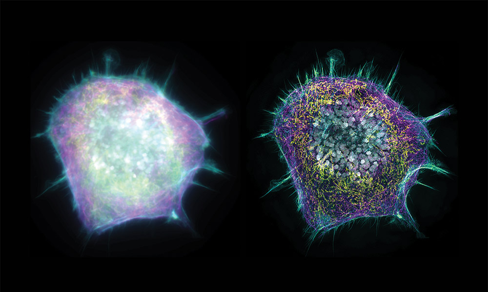
An image of multiple mitochondria, the powerhouse of the cell. Upper right corner shows a conventional microscope image, which is diffraction-limited to about 250 nm lateral resolution. The rest of the image shows results from dSTROM, a single molecule location microscopy technology capable of roughly 25 nm lateral structural resolution. Upper left inset is a direct comparison of the conventional microscopy (left) and super resolution microscopy (right). Photo credit: Markus Sauer, University of Wuerzburg.
Two super resolution fluorescence microscopy approaches formed the basis for the 2014 Nobel Prize in Chemistry: stimulated emission depletion (STED) and PALM, a single molecule localization microscopy technique. The Nobel committee said each technique transformed microscopes into nanoscopes.
As regards STED, it adds a second laser to a fluorescence microscope, with this additional high-intensity laser creating a donut-shaped depletion beam superimposed on a low-power laser excitation beam. The beam duo traverses the sample, gathering data. The excitation light turns the fluorophores on while the second laser shuts them down. Thus, the fluorophores only glow in the center of the beam area, and this arrangement of lasers, if pulsed in the right sequence, gets around the diffraction limit.
For this to work, the fluorophores must have the appropriate photochemistry. With STED, lateral resolution can be as fine as 40 nm. The theoretical lower limit is set by the depletion laser intensity, according to Lothar Schermelleh, a professor of bioimaging at Oxford who has co-authored papers that review SRM technologies. He adds that fluorophore photostability and other factors also play a role in effective resolution.
Researchers have used STED extensively, including Nobel Laureate Stefan Hell, who turned to STED to peer inside brain cells to better understand brain synapses. Researchers at the Max Planck Institute for Biophysical Chemistry deployed STED to image details inside mitochondria, the powerhouse of the cells, in living tissue, something impossible to do with standard microscopy because of the diffraction limit. The information gained can help in understanding what changes happen during aging. The research can’t be done with electron microscopy because it only works with fixed, dead samples.

Illustration: ©Johan Jarnestad/The Royal Swedish Academy of Sciences
Regarding single-molecule localization microscopy, there are a number of different approaches. One of the most popular is STORM—stochastic optical reconstruction microscopy—and it has variants, like Sauer’s contribution dSTORM. In discussing STORM and dSTORM, Sauer begins by noting that certain fluorophores blink. When excited, they glow for a while and then turn off, creating a random flickering of lights. Single-molecule localization microscopy exploits this response, using chemistry to create the right conditions for SRM.
“The off state is substantially longer [in time] than the on state,” Sauer says of how the chemically adjusted fluorophores blink.
So, the fluorophores briefly shine brightly and then go dark for a much, much longer period. It’s like a connect-the-dots picture, with the dots appearing separated by great distances, at random, and over time. By collecting enough of these points of light, systems produce data from which software generates an image. Microscopes can determine molecular location with a precision of about 10 nm.
Currently, researchers use single molecule localization microscopy to study nerve cells to see how synapses work—and to probe what goes wrong when they don’t function normally as is the case in epilepsy, Parkinson’s disease, or Huntington’s disease. Researchers also turn to super resolution to study proteins in healthy and diseased blood cells, enabling better understanding of the differences and mechanisms at work.
There are also combinations of the Nobel award-winning SRM methods.
Abberior Instruments, for instance, teams STED with single molecule localization microscopy to get determination of a molecule’s location down to 1 nm.
There are barriers, though, when it comes to translating this localization improvement to even better resolution. Sauer notes that in order to get a resolution of 20 nm, experience says there should be a signal from a fluorophore every 4 to 5 nm, half the minimum distance called for by information theory. That density requirement runs up against the fact that the biomolecular probes that bear the fluorophores are not infinitely small.
“We have to attach a fluorophore,” Sauer says. “And the probes, they all exhibit the size of a few nanometers, at least.”
Thus, the structural resolution of single molecule localization microscopy is today about 20 nm, largely because it isn’t possible to get a dense enough carpet of fluorophores on a specimen. However, there may be a solution emerging, one that involves a different concept entirely. It works by making things bigger, not by making resolution finer. Expansion microscopy is a brainchild of a team led by Edward Boyden of the Massachusetts Institute of Technology. It involves linking proteins in a biological specimen to the matrix of a hydrogel, the same stuff used in diapers to keep babies dry, and softening the tissue of the specimen by destroying the intermolecular interactions between the proteins.
The next step is to add water, causing the hydrogel to expand four-fold or more. If this transformation is even in all directions, then the relative positions of all the components of the tissue stay the same. So, a 20 nm resolution on the expanded specimen will be the same as a 2 or 1 nm resolution on the original sample, if the expansion is 10x or 20x. In this way, single-molecule localization microscopy can effectively rival the resolution possible with electron microscopy.
Combinations of expansion microscopy and SRM are the subjects of active research. Sauer thinks that expansion microscopy will revolutionize light microscopy, with one of the possibilities being the ability to expand single cells 100x or 200x so that they become visible to the naked eye.

Super resolution single molecule localization microscopy image of nuclear pore cell complexes, cellular structures that regulate biochemical flows into and out of the cell nucleus. At 100 nm diameter, they are too small for conventional microscopy to image clearly. Photo credit: Markus Sauer, University of Wuerzburg.
As for the state of today’s super resolution microscopy, Salvatore Finale, senior product manager at single molecule localization microscope maker ONI, notes that a 15 nm resolution class platform exists today with a large field of view, stable illumination, autofocus, and automation. Looking to the future, breakthroughs that would be useful in different photonic components could include highly sensitive, high-speed quantitative CMOS sensors with smaller pixels, he adds. Such sensors have extremely low noise with fast readout capabilities. They allow accurate measurement of the number of photons, enabling the discrimination of single photons within each pixel.
According to Finale, other desirable advances include stable and tunable high-power lasers, along with advanced adaptive optics to counteract optical aberrations and correct for imaging distortions deep within tissues. Finally, the development of brighter, more robust fluorescent probes is needed to complement these hardware improvements.
However, even though researchers would benefit from these innovations, they are more likely looking for better software and an easier-to-use system. “Most biologists do not want to assemble a super resolution microscopy stack. They want a reliable answer,” Finale says.
Regarding which SRM approach does that and is the best to use comes down to the scientific questions asked, Watkins says. The super resolution technique that helps researchers find an answer will vary. For instance, having to screen many different samples to look for specific fluorescently visible mutations may mean that a lower resolution, but faster system will be the right one. On the other hand, if a researcher is trying to approach electron microscopy performance, then only the highest resolution will do.
There is another issue to consider. Better and better resolution comes at a price. Finer resolution often means that there are fewer photons available over a given time period, which means it takes longer to capture a large enough number of photons. Also, typically the field of view at higher resolution is smaller, which increases the time needed to generate an image that spans a given area of a sample.
“Super resolution becomes really difficult because it takes such a long time to acquire the data and also to process the data,” Schermelleh says.
For a last point about the future of SRM, consider that the imaging center Watkins runs has 26 confocal microscopes and a few SRMs, with at least one representative of each of the different categories. The super resolution instruments are not used as much as the confocal systems, partly because the most advanced super resolution microscopes tend to be more complicated to learn to use and more difficult to operate.
From his experience, Watkins draws a conclusion about SRM and its future. “The very clear take home to me is that the easier it is to use, the more it penetrates.”
Hank Hogan is a freelance science and technology writer.