White matter pathways allow distant parts of the brain to communicate, supporting memory, emotion, and language. One such pathway, the uncinate fasciculus, connects the front of the temporal lobe with regions of the frontal cortex involved in decision-making and social behavior. Despite its importance, little is known about the microscopic structure of this tract in the human brain. Traditional techniques such as electron microscopy can reveal fine details, but they often fail when applied to postmortem human tissue, which is frequently degraded.
In a study published in Biophotonics Discovery, researchers report a new way to examine white matter structure in postmortem human brains. Using an advanced optical imaging method combined with deep learning, the team provides the first quantitative measurements of axon size and myelin thickness in the human uncinate fasciculus.
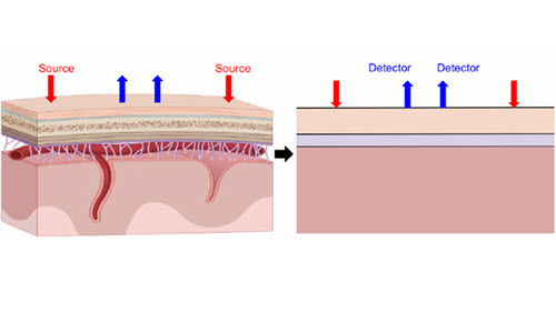
The study focused on myelin, the fatty insulating layer that surrounds many nerve fibers and allows electrical signals to travel quickly. Instead of relying on stains or labels, the researchers used spectral‑focusing coherent anti‑Stokes Raman spectroscopy (sf‑CARS). This technique detects natural molecular vibrations within tissue, making it possible to image lipid‑rich structures such as myelin directly.
Compared with older versions of CARS microscopy, the sf‑CARS system used in this study offers improved spectral resolution. The researchers achieved this by stretching ultrafast laser pulses with a glass rod, which sharpened the chemical signal from lipids and improved image contrast. Tests with reference materials showed that the modified system produced narrower spectral peaks and stronger signal overlap, confirming its suitability for myelin imaging.
Importantly, the method worked on human postmortem brain tissue with relatively long delays between death and fixation, which is a common limitation for brain bank samples.
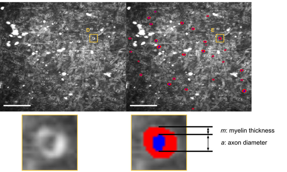
High‑resolution images alone are not enough to quantify brain structure at scale. To extract meaningful measurements, the team paired sf‑CARS imaging with a custom deep‑learning segmentation model based on AxonDeepSeg. The model was trained to distinguish axons and their surrounding myelin sheaths within microscopy images.
The researchers used an active learning strategy, gradually improving the model by correcting its predictions and retraining it with new data. This approach reduced the amount of manual annotation required while maintaining biological accuracy. After segmentation, automated quality control steps removed implausible structures, such as axons that were too small to measure reliably or had unrealistic geometry.
The final dataset included more than 2,600 myelinated axons from six individuals with no known psychiatric or neurodegenerative disorders.
Using this pipeline, the researchers measured axon diameter, myelin thickness, and the g‑ratio, a standard measure of how thick the myelin sheath is relative to the axon it surrounds.
They found that axon diameters in the temporal segment of the uncinate fasciculus ranged from about 0.4 to 6.4 micrometers, with an average diameter just under 1 micrometer. Myelin thickness averaged about half a micrometer. As expected from basic neurobiology, thicker axons tended to have thicker myelin, and the relationships between all measured features followed established patterns.
The team also compared the uncinate fasciculus with white matter from the anterior cingulate cortex in the same individual. Fibers in the uncinate fasciculus had thicker myelin and lower g‑ratios, consistent with the needs of long‑range connections that transmit signals rapidly over greater distances.
This work provides the first direct measurements of ultrastructural features in the human uncinate fasciculus and establishes a method that can be applied to other brain regions. Because the approach does not require special stains or near‑perfect tissue preservation, it opens the door to studying large collections of postmortem human brains that were previously inaccessible to ultrastructural analysis.
The authors emphasize that the method currently detects only myelinated axons and works best when fibers are oriented favorably within tissue sections. Still, combining sf‑CARS imaging with automated segmentation allows researchers to analyze far more axons than manual methods would permit.
In the future, this pipeline could be used to compare white matter structure across brain regions, between hemispheres, or between healthy tissue and brains affected by psychiatric or neurological disorders. By making microscopic measurements feasible in human postmortem samples, the study provides a practical foundation for linking brain structure to behavior and disease.
For details, see the original Gold Open Access article by K. Perlman, V. P. Noel, A. Collin, et al., “Ultrastructural analysis of human uncinate fasciculus with coherent anti-Stokes Raman spectroscopy,” Biophoton. Discovery 3(2), 025002 (2026), doi: 10.1117/1.BIOS.3.2.025002