On the edge of Tokyo, at Japan’s national research institute RIKEN, a junior researcher, Yuya Morimoto, and his ultrashort electron beam science group are preparing to observe what was once thought unobservable—how individual electrons move within a single molecule. As a Hakubi Fellow—a title awarded to “exceptionally talented individuals”—Morimoto hopes to capture the infinitesimally fleeting motions of these charged particles as they migrate, tunnel, or hop during ultrafast chemical processes in proteins, DNA, and more. To do so, he needs to build the fastest electron microscope in the world: an attosecond electron microscope.
“The electrons within our molecules react at the atomic scale and on the attosecond timescale, so that’s why I’m developing attosecond electron microscopy here in Japan,” says Morimoto. “If we can see this kind of electron motion, then we will understand so much more about our bodies and life.”
The attosecond electron microscope lives up to its name because it is designed to observe phenomena that play out at attosecond—one quintillionth of a second—timescales. To put this flicker of a second into perspective, there are approximately as many attoseconds in one second as there are seconds in 13.8 billion years—the age of the universe. But so far, only a handful of attosecond electron microscopes exist. Mohammed Hassan, who heads up the Attomicroscopy and Ultrafast Quantum Imaging Center at the University of Arizona (UA), built one such instrument. As he puts it: “To see electron motion in real time and space, you have to find a way to bring attosecond resolution to the electron microscope, and this has been very difficult for everyone.”
Still, Hassan made science headlines last year when, using ultrafast electron diffraction, his group recorded movies of electrons moving through a sheet of graphene. The researchers had pushed their attosecond electron microscope to reach a temporal resolution—the smallest time interval that can be captured between consecutive images—of just 625 attoseconds. Hassan claimed a Guinness World Record for the fastest electron microscope but, like electron motion, this distinction could be fleeting.

Professor Mohammed Hassan leads the Attomicroscopy and Ultrafast Quantum Imaging Center at the University of Arizona. Photo credits: Mohammed Hassan/University of Arizona.
Attosecond electron microscopy follows more than two decades of development of ultrafast electron microscopy based on the transmission electron microscope (TEM), which uses a continuous stream of electrons to image atomic structures as small as 0.05 nm, or half an angstrom. But the images generated are static. When it comes to capturing the dynamics of nanoscale molecules and materials, a TEM instrument is held back by its detector’s video-recording rate, which limits temporal resolution to milliseconds. So, while TEM can track fast processes such as charge transfer at battery electrodes or defects moving in semiconductors, science’s most transient processes, including electron motion, have remained elusive.
To overcome TEM detectors’ recording rate, researchers, including the late Ahmed Zewail of Caltech who won the 1999 Nobel Prize in Chemistry, started to integrate ultrafast lasers. Zewail had used ultrashort laser flashes to investigate how atoms move in a chemical reaction—a field dubbed femtochemistry—and wanted to apply this concept to TEM to create ultrafast electron microscopy.
By 2008, Zewail and Caltech colleagues had constructed an ultrafast TEM with sub-picosecond—less than one trillionth of a second—temporal resolution and imaged atomic-scale motions in gold nanostructures and graphite nanosheets. The instrument used a pump-probe laser setup in which a pulsed femtosecond laser—with a pulse duration of one quadrillionth of a second—was directed at the sample to trigger ultrafast dynamics. Meanwhile, a second pulsed laser illuminated the photocathode in the TEM’s electron gun, generating picosecond pulses of electrons to interact with the sample and form an image.
With this setup, sequential images, generated by millions of electrons, could be recorded and assembled to create atomic-scale movies down to a few hundred femtoseconds resolution. Critically, temporal resolution now depended on the duration of the electron pulses, rather than the detector’s relatively slow recording rate. Next, Zewail and colleagues unveiled photon-induced near-field electron microscopy (PINEM), a form of ultrafast electron microscopy that would become the bedrock of attosecond electron microscopy. They fired their TEM’s electron pulses at the rapidly oscillating fields of light around carbon and silver nanostructures that were being pumped and excited with the ultrafast laser pulses. By overlapping the electron pulses, the laser pulses, and the nanostructures in this way, the researchers could use the TEM to explore the manically fast, light-matter interactions playing out in the optical field.
Zewail and many other scientists went on to exploit this PINEM effect to reshape TEM electron pulses into ever-shorter bursts, pushing back the boundaries of ultrafast electron microscopy. Remarkable imagery of ultrafast processes emerged, including crystallization, melting, plasma waves evolving, and much more. However, along the way, a handful of scientists were also working on the next temporal frontier: attosecond resolution.
Attosecond electron microscopy development has been led by the UA’s Hassan, as well as Peter Baum, head of the Light and Matter Group at the University of Konstanz, and Claus Ropers, head of Ultrafast Dynamics and director at the Max Planck Institute for Multidisciplinary Sciences. Baum and Hassan both spent portions of their time as postdoctoral researchers with Zewail at Caltech, but across the last decade or so, each researcher separately has developed PINEM-based ways to get their TEMs to beat to an attosecond tempo.
By early 2024, each had generated remarkable videos of light-matter interactions with their attosecond electron microscopes, claiming “the world’s fastest electron microscope” within months of one another. Baum and colleagues used the rapid light oscillations of a continuous wave laser to bunch the electron beam into a sequence of attosecond pulses and created movies of chiral surface waves emerging in a tungsten nano-needle tip. Morimoto worked with Baum on this before moving to RIKEN in 2021. Meanwhile, Ropers and colleagues measured, with attosecond resolution, changes in how electrons behave when they interact with laser light fields. By precisely controlling this interaction, they imaged the optical field oscillations within gold nanoprisms. In hot pursuit of electron motion, Hassan and colleagues used the optical field from an ultrashort laser pulse to select, or gate, a section of the electron pulse. Using their gating approach, they isolated just a single electron with an electron pulse that repeated every 625 attoseconds and imaged electrons moving through a graphene sheet. They were recognized as having the world’s fastest electron microscope in August 2024.
In November 2024, Baum and Ropers wrote a comment for Science Advances—which had published Hassan’s results—highlighting what they believe to be experimental inconsistencies in Hassan’s work that render his claims and conclusions unsubstantiated. Hassan countered, providing experimental evidence and clarifications, and challenging the other approaches.
As both sides stand their ground, Hassan reflects: “I always wanted to learn how the electron moves…and built my lab around optical gating and the attosecond electron microscope to do this. With our microscope, we hope the scientific community can understand the quantum physics behind how an electron behaves and moves.”

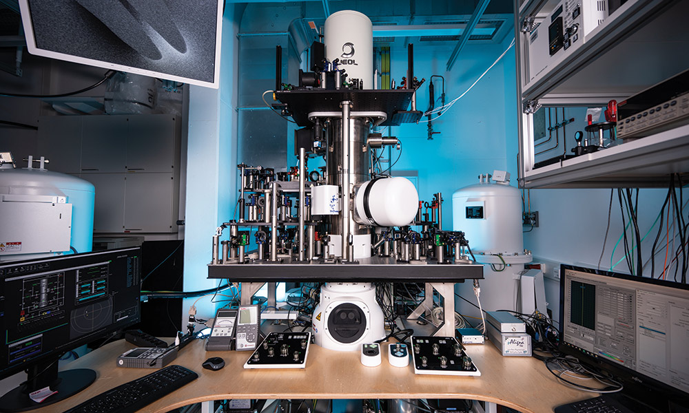
Doctoral student John Gaida at the attosecond electron microscope in the Ultrafast Dynamics group at the Max Planck Institute for Multidisciplinary Sciences. Photo Credit: Irene Böttcher-Gajewski / Max Planck Institute for Multidisciplinary Sciences.
As world records are broken, accessing an ultrafast or attosecond electron microscope hasn’t been easy. Tom Gage and colleagues at Argonne National Laboratory’s (ANL) Center for Nanoscale Materials noted this back in 2019, and so set to modifying their TEM with an ultrafast laser system. By 2021, their ultrafast electron microscope was unveiled, making ANL the first national user facility to offer such an instrument, and studies of short-lived phenomena have ensued.
Taking the temporal resolution of the ultrafast TEM to the attosecond level is on Gage’s radar, but as he puts it: “While we’re kind of dipping our toe in the water here, it’s a fairly large hardware endeavor and it’s not easy.” But change is afoot.
Recent industry developments indicate that more straightforward ultrafast, and even attosecond, electron microscopy could be coming. For example, the Max Planck Institute for Multidisciplinary Sciences recently licensed an ultrafast TEM electron source, developed by Ropers and colleagues, to electron microscope manufacturer JEOL and its subsidiary, Integrated Dynamic Electron Solutions (IDES). Ropers is confident commercial availability of these technologies will drive ultrafast science forward. And as IDES Chief Technology Officer Bryan Reed, puts it: “If you ever wanted to play with ultrafast light-matter interactions at the atomic scale, now’s your chance.” Baum would like to see the attosecond electron microscope one day become a standard tool. “We have a few patents [for attosecond technology] and are in discussions with several electron microscope manufacturers from around the world,” he says. “Our modifications are actually rather simple…. We hope to create a commercial attosecond electron microscope that researchers and engineers can just purchase and apply.” Still, he cautions, this could take place between one to five years from now, or never.
Hassan has also patented his system and method as “attomicroscopy:” attosecond electron imaging and microscopy. But perhaps the most immediate indication of broader tech adoption is a primer on ultrafast electron microscopy being written by Fabrizio Carbone and colleagues at École Polytechnique Fédérale de Lausanne’s Laboratory for Ultrafast Microscopy and Electron Scattering. Carbone was at Caltech when the first ultrafast TEM was developed, and he worked with Zewail on implementing femtosecond spectroscopy in an electron microscope in 2009. As he explains, the primer will help researchers develop and use the technology, and he expects readers will range from PhD students to research group leaders.
“More and more researchers are trying to get ultrafast microscopy into their lab, but it’s a complicated technique as you need to be an expert in both ultrafast lasers and the transmission electron microscope,” he says. “We want to smooth this learning curve of ultrafast electron microscopy.”

Temporal evolution of a light field at a gold nanoprism. The time between the images is 640 attoseconds. Photo credit: John Gaida / Max Planck Institute for Multidisciplinary Sciences.
So as ultrafast electron microscopy edges towards more widespread use, where will the method take science next? Whilst the pursuit of observing ever-more arcane light-matter interactions and electron motion in materials continues, pushing this to the quantum realm is already happening.
Ropers is excited about eventually using ultrafast and attosecond electron microscopy to investigate dynamic processes within quantum dots, superconducting qubits, and other individual quantum systems. He and colleagues are experimenting with entangling electrons with photons with a view to interrogating quantum phenomena. “How do you access quantum fluctuations and correlations? How do you probe entanglement? How do you trace decoherence?” he asks. “This is long-term research, but we and many international colleagues are working on the solutions.”
Hassan and his team are keen to study the quantum properties of electrons moving in different materials and are working on a quantum squeezed light source to drive the electron dynamics. “It’s good to image electrons but we also want to demonstrate the uniqueness of the attomicroscope, and I think one way to do this is to study quantum electron dynamics with attosecond resolution,” he says. “We are knocking on a door that nobody has passed through yet, and still need more technical development…. But this could unlock possibilities for quantum technologies using ultrafast quantum light, including petahertz-scale secure quantum communication, quantum computing, and ultrafast spectroscopy.”
Hassan also has the ambitious goal of one day building an ultrafast quantum imaging center that includes your “everyday” attosecond electron microscope, a quantum version of that, and even a cryogenic attosecond electron microscope to probe structures with unprecedented precision. Indeed, for Hassan, the future is not just about quantum physics. “We want to go quantum biology,” he says. “This is a new field, and we want to prove how important electron dynamics and quantum behavior are to biological processes.”
As a starting point, Hassan is working on electricity-conducting cable bacteria with Moh-El-Naggar at the University of Southern California. In 2012, El-Naggar showed these electric marvels can transport electrons centimeter distances along their length, and now Hassan intends to explore the electron behavior. Experiments on desiccated samples are underway, with studies on living bacteria in liquid-cell holders to follow.
“A key motivation is to replace the normal wires in medical devices with cable bacteria, which will be more compatible with the body,” says Hassan.
In the future, Hassan hopes his ultrafast quantum imaging center will be used to scrutinize more biological phenomena, including electron migration in proteins and electron tunneling in DNA—a vision aligned with that of RIKEN’s Morimoto. The Hakubi Fellow will initially study micro- or nanocrystals of biomolecules, and he hopes to have his attosecond electron microscope, which will use the same electron pulse compression scheme as Baum’s instrument, up and running in the next year or so.
But there’s more: Morimoto is also excited about seeing ultrafast electron microscopy reach the next temporal resolution regime, the zeptosecond, which at one trillionth of a billionth of a second is the smallest unit of time ever measured. Realizing an instrument that can capture transient phenomena that linger for only the briefest moment of time seems truly remarkable, but according to the researcher this could happen sooner than you’d think.
“Maybe in 10 years we could get into this zeptosecond regime,” Morimoto says. “I think researchers will want to go there as a lot of attosecond physics is going to be investigated by the electron microscope.”
Morimoto also has a few ideas of what could be explored. Nuclear fusion power plants could well emerge in the next decade, and he believes researchers will want such a tool to investigate fusion. He also points to the burgeoning field of nuclear clocks that, once realized, will use the vibrations of atomic nuclei to keep exquisitely precise time.
“Scientists across Europe, the US, and Japan are working hard on nuclear clocks, and would be able to investigate their dynamics [with a zeptosecond microscope],” he says. “I can’t really comment on what is hidden or unknown—but it will be good to see what’s going on inside the nucleus.”
Rebecca Pool is a science and technology journalist based in the UK.